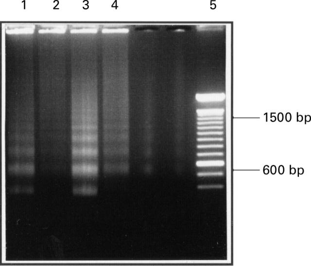
Figure 3 .
Fragmentation pattern of low molecular weight DNA as revealed by electrophoresis on agarose. Cells were treated with 25 µM C6-ceramide in the presence (lanes 2 and 4) and absence (lanes 1 and 3) of 1 mM S-nitroso- N-acetyl-penicillamine. Extracts from cells attached to the culture plate are in lanes 1 and 2 and cells that had detached are in lanes 3 and 4. Markers are in lane 5.

