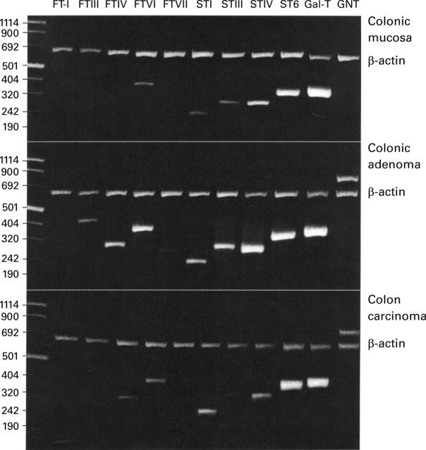
Figure 1 .
Glycosyltransferase expression in human colorectal tissue from case 3. Fluorescence of electrophoretically resolved, ethidium bromide stained polymerase chain reaction (PCR) products of β-actin and glycosyltransferase target sequences from colon mucosa, colon adenoma tissue, and colon carcinoma tissue is shown. Glycosyltransferases were amplified as described in Materials and methods. The upper band shows the reaction product of β-actin amplification. All amplicons were sequenced and compared with published sequences. First lane, molecular mass markers (bp). FT-I, H blood group α1,2-fucosyltransferase I; FTIII-VII, fucosyltransferases III-VII; STI-IV, sialyltransferases ST3Gal-I-IV; ST6, sialyltransferase ST6Gal-I; Gal-T, β1,4-galactosyltransferase; GNT, β1,6-N-acetylglucosaminyltransferase V.

