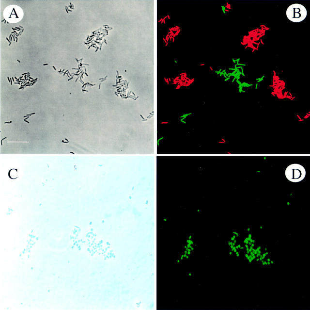
Figure 2 .
Specific detection of clarithromycin sensitive and resistant H pylori isolates. (A) Phase contrast microscopy of a mixture of clarithromycin sensitive and resistant H pylori isolates. (B) The same microscopic field after hybridisation with probes ClaWT-FLUOS (green) and ClaR1-Cy3 (red). (C) Phase contrast micrograph showing coccoid forms of clarithromycin resistant H pylori. (D) Whole cell hybridisation of coccoid forms with probe Hpy-1-FLUOS. Bar represents 10 µm.

