
Figure 1 .
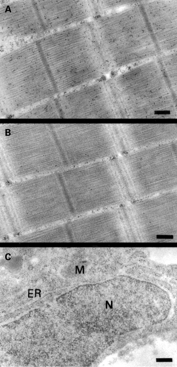
Subcellular localisation of parvalbumin in carp muscle and liver by immunogold electron microscopy. Bright field micrograph of ultrathin sections of carp muscle labelled with a monoclonal antibody against parvalbumin (A) and with an isotype matched control antibody without specificity for parvalbumin (B). (C) A section from carp liver labelled with the monoclonal antibody against parvalbumin. ER, endoplasmic reticulum, M, mitochondria, and N, nucleus. Magnification in all micrographs is 58 000 ×. The bars correspond to 0.25 µm. Black dots represent bound gold particles.