
Figure 3 .
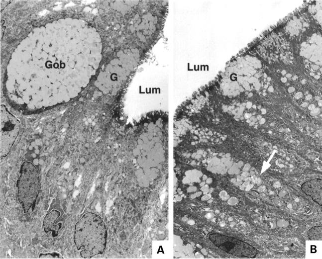
Electron micrograph of two areas within a patch of focal metaplasia. In each, Lum=lumenal surface. (A) Gob, a cell containing mucous secretory granules typical of intestinal goblet cells; G, mucus secretory granules typical of gastric foveolar cells. (B) Arrow shows single cell containing granules typical of both gastric and intestinal phenotypes; G, mucus granules with gastric phenotype.