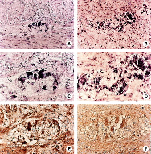
Figure 4 .

Localisation of neurokinin-1 receptor (NK-1R) expression in non-inflamed (A, C, and E), and inflamed Crohn's disease (CD) (B, D, F) myenteric plexus by in situ hybridisation (A-D), and immunohistochemistry (E, F). (A) NK-1R mRNA (purple) is detected in enteric neurones of the myenteric plexus of normal colon. (C) Higher magnification view of (A). (E) NK-1R protein (brown) staining is evident in the myenteric plexus of normal colon. Section was counterstained with haematoxylin (blue). (B) Expression of NK-1R mRNA by enteric neurones of the myenteric plexus of CD colon. Note the NK-1R positive cells of lymphoid morphology. (D) Higher magnification view of (B). (F) Immunohistochemical detection of NK-1R protein expression (brown) in the myenteric plexus of CD colon. Section was counterstained with haematoxylin (blue).
