
Figure 3 .
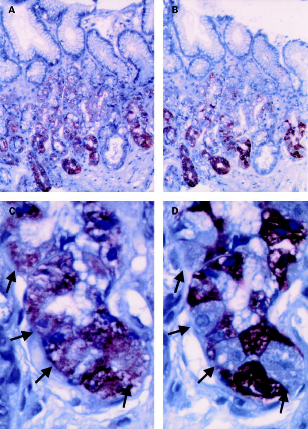
Immunohistochemical detection of leptin (A, C) and pepsinogen (B, D) in serial sections of human fundic mucosal biopsies. (A, B) Overview (original magnification ×100). (C, D) Detailed view (original magnification ×400). Arrows denote corresponding cells of adjacent sections which are leptin positive and pepsinogen negative. Paraffin embedded, formalin fixed tissue sections were analysed using a streptavidin-peroxidase technique as described under materials and methods.