
Figure 5 .
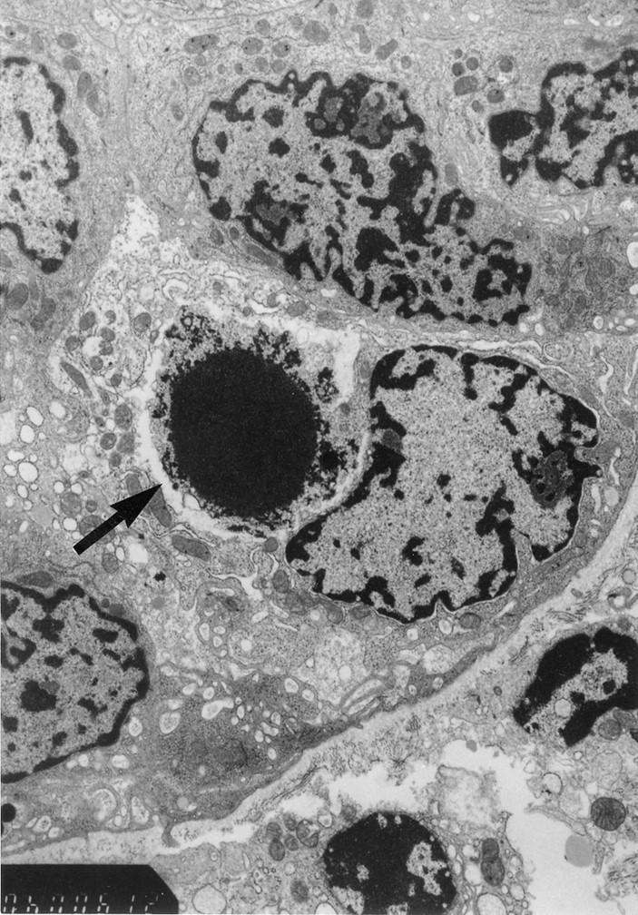
Electron micrograph of an apoptotic cell in the epithelial layer of the small intestinal crypt at one day after chemotherapy. The arrow indicates an apoptotic cell with a pyknotic nucleus. (Original magnification ×12 000.)
Official websites use .gov
A
.gov website belongs to an official
government organization in the United States.
Secure .gov websites use HTTPS
A lock (
) or https:// means you've safely
connected to the .gov website. Share sensitive
information only on official, secure websites.
Electron micrograph of an apoptotic cell in the epithelial layer of the small intestinal crypt at one day after chemotherapy. The arrow indicates an apoptotic cell with a pyknotic nucleus. (Original magnification ×12 000.)