
Figure 3 .
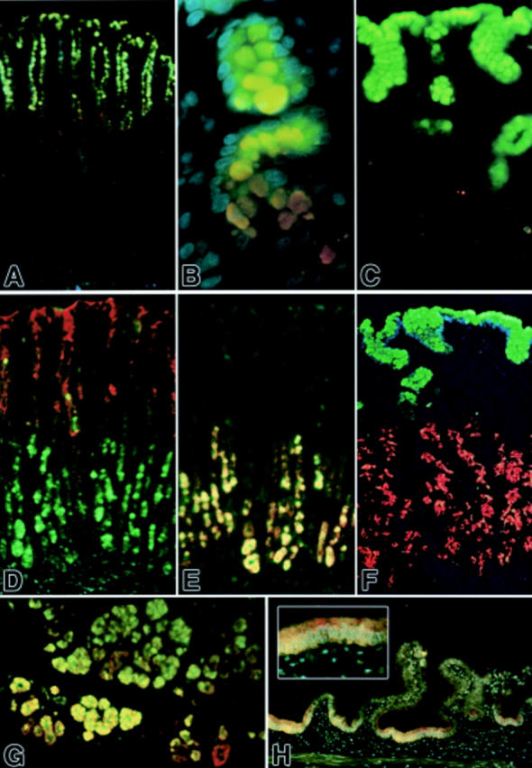
Fluorescent immunohistochemical localisation of mucin glycoproteins and trefoil factor family (TFF) peptides in the stomach, duodenum, and gall bladder. (A, B) TFF1 (green) and MUC5AC (red) colocalised in the gastric surface epithelium; MUC5AC immunoreactivity continues deeper into the isthmus and neck region (DAPI counterstain). (C) TFF1 (green) appears abundant in gastric foveolar surface cells whereas MUC5B (red) appears weakly localised to some mucous neck cells (weak DAPI counterstain). (D) TFF2 (green) and MUC5AC (red) were mostly segregated, with TFF2 immunoreactivity abundant in basal parts of these gastric glands and MUC5AC glycoprotein abundant in the epithelium at the surface, isthmus, and neck. Colocalisation was infrequent (DAPI counterstain). (E) TFF2 (green) and MUC6 glycoprotein (red) were mostly colocalised and confined to the basal parts of these gastric glands. (F) TFF1 (green) and MUC6 glycoprotein (red) were mostly segregated, with TFF1 abundant in surface epithelium and MUC6 appearing confined to epithelium in the lower half of these gastric glands, including some unequivocal mucous neck cells (red; upper left) (DAPI counterstain). (G) TFF2 (green) and MUC6 glycoprotein (red) were abundant in epithelium in the acini of Brunner's glands of the duodenum, and are most often colocalised (weak DAPI counterstain). (H) TFF2 (green) and MUC6 glycoprotein (red) appear colocalised (orange-yellow) in some epithelium of this non-inflamed gall bladder. Inset shows enlargement of central region (DAPI counterstain). Original magnifications: A, D, E, H 100×; B 400×; C 250×; F 200×.