
Figure 6 .
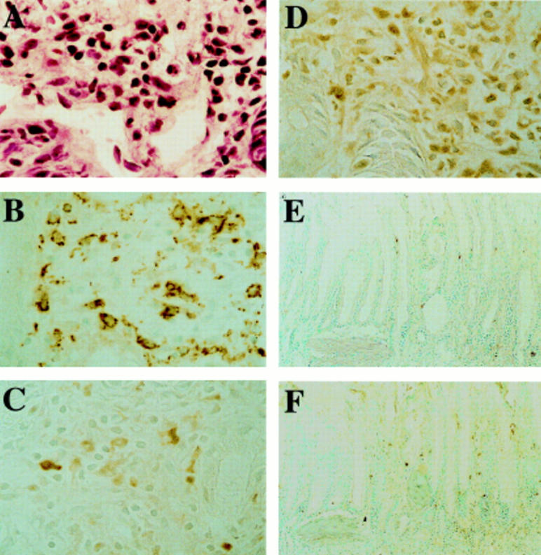
Cellular infiltration and immunohistochemical staining for interleukin 1β (IL-1β) and tumour necrosis factor α (TNF-α) in superficial mucosa at 24 hours after IL-1β treatment. (A) IL-1β induced infiltration by leucocytes, including neutrophils, in the superficial portion of scarred mucosa. (B) Monocytes/macrophages were abundant in this region. IL-1β (C) and TNF-α (D) were detected mainly in inflammatory cells in the superficial mucosa. The numbers of cells stained for IL-1β (E) and TNF-α (F) were small in rats given the antibody against intercellular adhesion molecule 1. Original magnification ×200 (A-D) and ×50 (E, F).