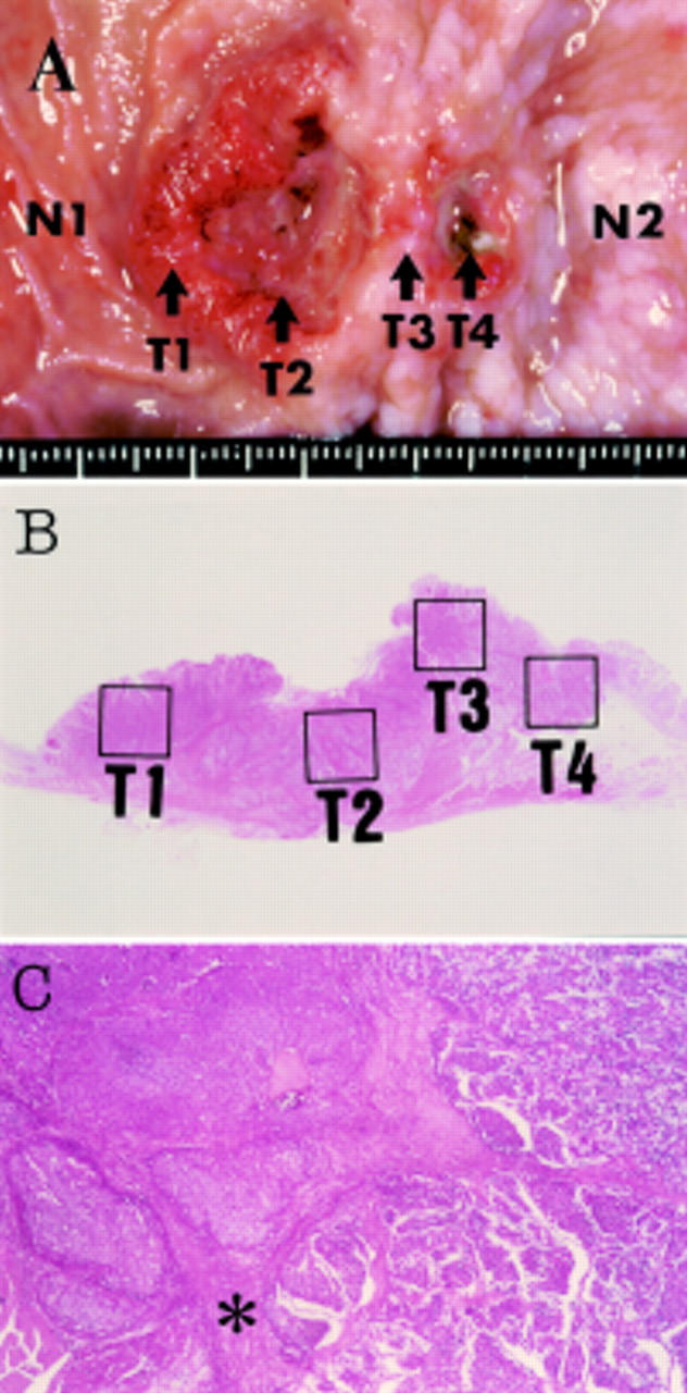
Figure 1 .

(A) Macroscopic view of the resected specimen showing a spherical tumour located at the pre-pylorus and duodenum. Tissues from six parts in and around the gastroduodenal carcinoma were microdissected to analyse p53 gene mutations: normal duodenum (N1), anal side of duodenal tumour (T1), centre of duodenal tumour (T2), tumour at the pyloric ring (T3), tumour of the stomach (T4), and normal stomach (N2). (B) Distinct areas (T1-T4) which were analysed for p53 gene mutation are indicated by the solid lines. (C) The well circumscribed tumour cell nests in the duodenal region revealed typical medullar growth of neuroendocrine carcinoma (left side). Poorly differentiated adenocarcinoma in the stomach showed small acinar pattern (right side). The pyloric sphincter (asterisk) clearly divides the two histologically distinct lesions.
