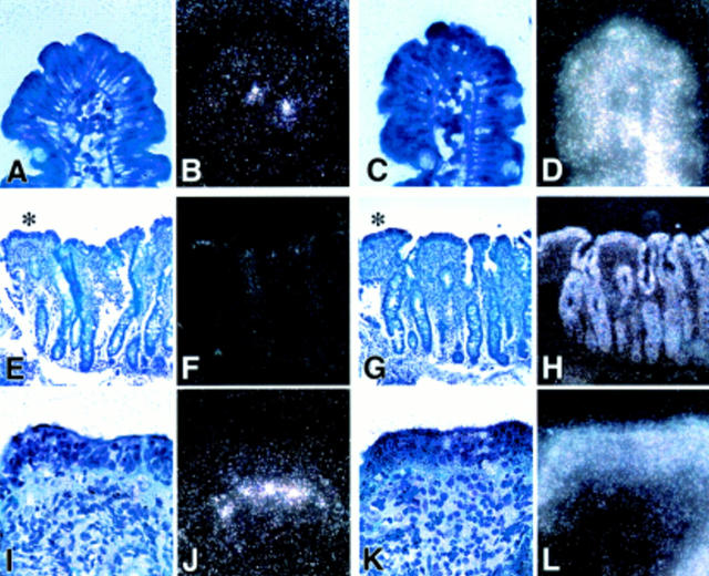
Figure 2 .
Detection of keratinocyte growth factor (KGF) mRNA by in situ hybridisation. (A, B) and (C, D) represent light/dark images of a control section hybridised with antisense probes to KGF mRNA (A, B) and to β-actin (C, D) as a positive control at a magnification of 50×. Very few KGF transcripts were detectable in control sections below the tip of the villi. (E) and (F) show light and dark field images, respectively, at 20× magnification stained for KGF. In (I) and (J) at 50× magnification the flat villi indicated by an asterisk in (E) are shown in more detail. KGF positive hybridising cells are distributed in the subepithelial region of the lamina propria below the tip of the flattened villi. (G, H) (20×) and (K, L) (50×) represent light (G, K) and dark field (H, L) images of the corresponding β-actin staining as a positive control.

