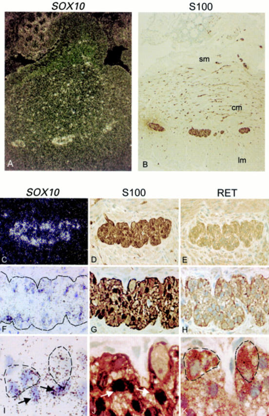
Figure 1 .

Expression of SOX10 in both neurones and glia of enteric plexuses in the colon of normal infants. (A) Dark field illumination of a cross section of normal colon showing strong in situ hybridisation signals in myenteric plexuses and in the nerves in the longitudinal and circular muscular layers. (B) Bright field illumination of an adjacent section showing S100 immunoreactivity in the SOX10 positive cells. (C) Dark field illumination and (F, I) bright field illumination of a myenteric plexus hybridised in situ with a SOX10 probe. (D, G, J) Immunohistochemistry of an adjacent section with S100 antibody showing that the glial cells present in the plexus are stained. (E, H, K) Immunohistochemistry of another adjacent section with RET antibody showing that the neurones and nerves in the plexus are stained. For comparison, in I, J, and K, the same glial cells are marked with arrows and the neurones are circled by broken lines. Both glial cells immunoreactive for S100 and neurones immunoreactive for RET had positive SOX10 hybridisation signals. Original magnification: (A, B) ×100; (C, D, E) ×400; (F, G, H) ×500; (I, J, K) ×1000. Sm, submucosa; cm, circular muscle; lm, longitudinal muscle.
