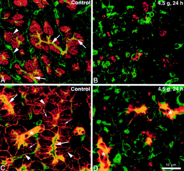
Figure 4 .
Effects of arginine injection on the distribution of amylase, cytokeratin, and actin. Cryostat sections from control animals (A, C) and in animals 24 hours post-injection (4.5 g/kg arginine) (B, D) were double labelled with BODIPY FL labelled phallacidin, and either antiamylase or anticytokeratin 8/18 antisera, followed by Cy3 conjugated second antibody. Immunofluorescence was monitored by confocal microscopy, and digitised images of FITC and Cy3 channels were superimposed. In control tissue (A), amylase (red fluorescence) is present in the secretory granules that fill the apical cytoplasm of acinar cells beneath the actin rich terminal web and lumen (arrows). Actin (green fluorescence) is also associated with basolateral membranes (arrowheads). Cytokeratin distribution (red fluorescence) in control acinar cells (C) is also present within the terminal web (large arrows) where it partially overlaps actin (yellow fluorescence) and along basolateral membranes (arrowheads). A thin band of cytokeratin free actin is present immediately beneath the luminal membrane (green fluorescence). In addition, filamentous cytokeratin strands extend in a rich network from the terminal region towards the basal aspects of the acinar cells (small arrows). Small ducts (asterisk) and their centroacinar cell termini express actin and abundant cytokeratin which overlap in their distribution. After 24 hours, the highly polarised distribution of actin, amylase, and cytokeratin in acinar cells is severely disrupted (B, D). Only a few small clusters of amylase containing secretory granules remain (B), and filamentous cytokeratin is lost, being replaced by a few focal deposits or small aggregates (D). Ducts and centroacinar cells (asterisk) retain actin and cytokeratin staining but in a markedly disorganised form. (A) and (B) are images reconstituted from complete Z series (1 µm steps) using Noran Intervision software. (C) and (D) are single optical sections taken at the focal plane in the Z series where both actin and cytokeratin filaments were defined most sharply.

