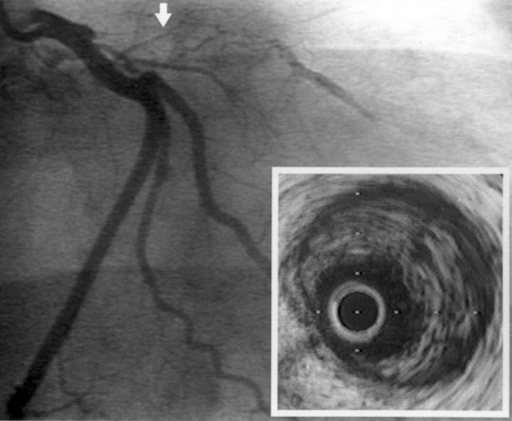
Figure 1 .
Coronary angiogram in the right anterior oblique projection of case 2 showing proximal occlusion of the left anterior descending artery and decreased filling distally. Inset, intravascular ultrasound image of the proximal left anterior descending artery (site indicated by arrow) showing extensive intraluminal thrombus.

