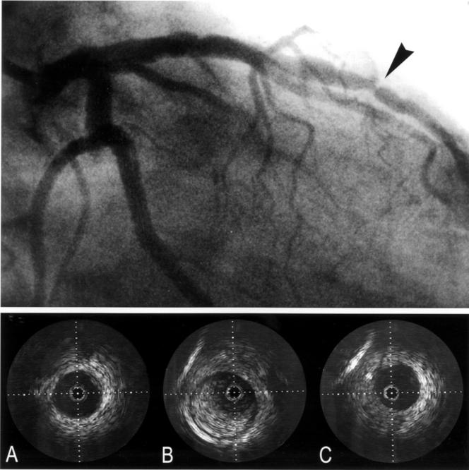
Figure 2 .
Angiogram and intravascular ultrasound images of the left anterior descending coronary artery in another patient. The arrow indicates the lesion. The intravascular ultrasound images show the proximal reference (A), lesion (B), and distal reference (C) site. At the lesion site arterial compensatory enlargement is present.

