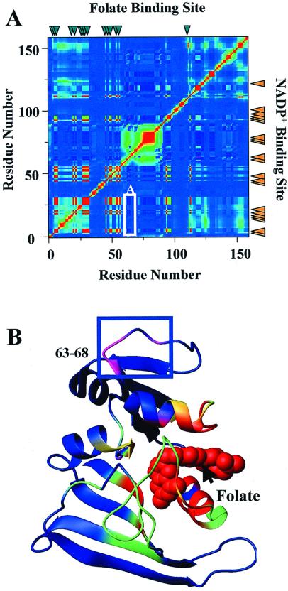
Figure 5.
FCs of the folate site. (A) FCs for the folate-binding site represented as described in Fig. 4 for the NADPH site. (B) High-resolution structure of DHFR (19) with van der Waals' surface of folate in its respective binding site. The negative FCs between the folate site and residues 64–68 (labeled A) are highlighted in B. Note the absence of a propagation pathway from the folate site. This figure was prepared by using the program molmol (38).

