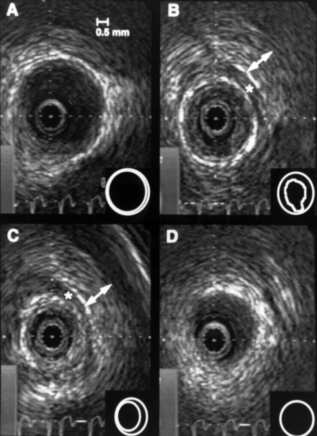
Figure 2 .
Intracoronary ultrasound images of the patient after PTCA of the left anterior descending artery within the myocardial bridge. A to D represent the course of the left anterior descending artery from proximal to distal. B and C are located within the myocardial bridge. A soft atheromatous plaque was detected that was not apparent on the coronary angiogram. Plaque rupture (B) was probably the result of the PTCA procedure. Asterisks indicate an echolucent band surrounding the artery within the bridge that probably represents adipose tissue around the artery. Vessel lumen and media bounded area are indicated in the insets, and plaque rupture can be seen in panel B at 6 o'clock.

