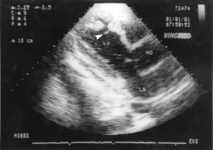
Figure 1 .
Four weeks after the onset of Dressler's syndrome (eight weeks after myocardial infarction) the pseudoaneurysm was primarily detected. Echocardiographic three chamber view shows perforation of the left ventricular wall, extracardial space, and floating parts of myocardial tissue at the margins of the perforation (arrow). LA, left atrium; LV, left ventricle; AO, aorta.

