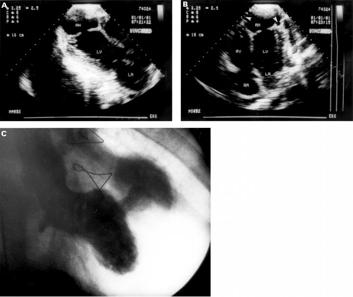
Figure 2 .
Six weeks later (14 weeks after myocardial infarction). (A) Three chamber view shows pericardial effusion, significant enlargement of the perforation, and of the false lumen. (B) Four chamber view shows echo-free edges at the margins of the false lumen (arrows), which was interpreted as progressive detachment of the pasted epicardium and pericardium. (C) Left ventriculography performed preoperatively (right anterior oblique projection) showing the large size of the pseudoaneurysm. LA, left atrium; RA, right atrium; RV, right ventricle; LV, left ventricle; AO, aorta; AN, false lumen of pseudoaneurysm.

