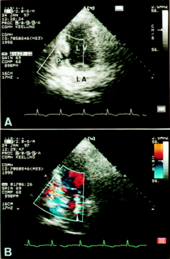
Figure 2 .

(A) Apical two chamber view of transthoracic cross sectional echocardiography showing an echo free space (pseudoaneurysm) posterior to the left ventricle. (B) Colour Doppler study demonstrating a flow entering the pseudoaneurysm (arrow) and a mitral regurgitant jet (arrowhead). LA, left atrium; LV, left ventricle; Ps, pseudoaneurysm.
