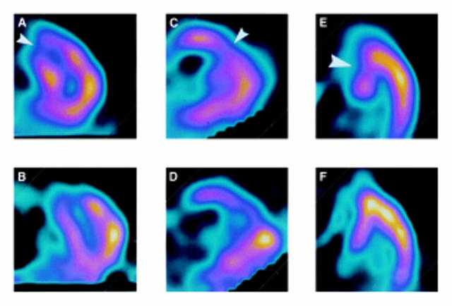
Figure 2 .
Sestamibi scan of patient 5 showing a fixed defect (indicating infarction) in the anterior wall (small arrowheads) and reversible ischaemia in the septum (large arrowhead). Transaxial views at mid-ventricular to basal level at (A) stress and (B) rest. Vertical long axis views at (C) stress and (D) rest. Horizontal long axis views at stress (E) and rest (F).(Refer to fig 1 for diagram of standard views).

