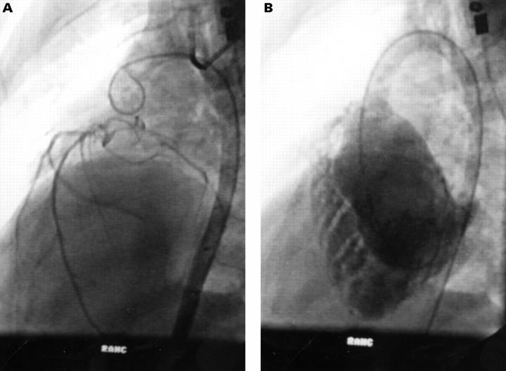
Figure 3 .
Angiograms of a (non-study) patient with congenitally corrected TGA. (A) Late phase aortogram (left anterior oblique view) showing inverted coronary artery pattern with sparse coronary supply to the right ventricle. (B) Right ventriculogram (left anterior oblique view) showing gross hypertrophy and trabeculation, especially of the apical half of the ventricle.

