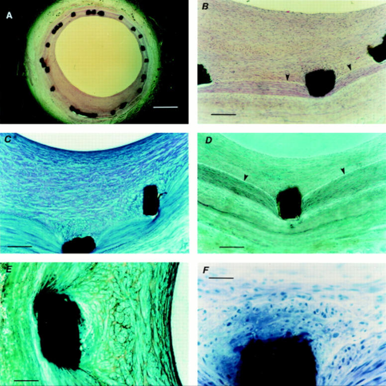
Figure 1 .

Histological staining of stented arterial sections. (A) Haematoxylin and eosin stained section (scale bar = 500 µm) showing preserved arterial wall architecture and stent struts in situ; and (B) at higher magnification (scale bar = 100 µm) with an intact internal elastic lamina (arrowheads). (C) Toluidine blue stained section (scale bar = 100 µm) showing varying spatial orientation of cells within the neointimal layer. (D) Trichromatic stained section (scale bar = 100 µm) showing struts in situ, preserved arterial architecture, and an intact internal elastic lamina (arrowheads); and (E) at higher magnification (scale bar = 50 µm) showing cells around the strut arranged in clusters, unlike cells nearer the lumen. (F) Haematoxylin stained section (scale bar = 5 µm) showing an intact tissue metal interface with neointimal cells around the strut arranged in clusters.
