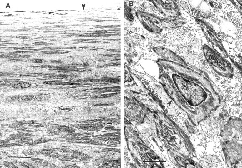
Figure 3 .

Transmission electron micrographs of stented coronary artery sections. (A) Neointimal layer 28 days after stenting with abundant extracellular matrix and interspersed smooth muscle cells (scale bar = 0.5 µm) arranged in distinctly different spatial orientation. Arrowhead indicates the luminal surface. (B) Phenotype of the neointimal cells is confirmed as smooth muscle cells (scale bar = 0.18 µm). Note characteristic cytoplasmic motor regions of these cells (arrowhead).
