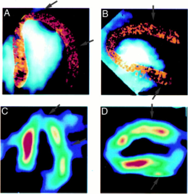
Figure 2 .
Examples of perfusion defects from two patients using apical four chamber (left panels) and apical two chamber (right panels) views. The top panels depict images obtained using MCE and the bottom panels show those obtained using SPECT. The MCE image from the apical two chamber view in this patient is placed on its side to correspond to the vertical two chamber view using SPECT. The first patient (left panels) had a large lateral defect that was identical on MCE and SPECT. The second patient (right panels) had vascular defects in the anteroapical and the upper posterior walls that were identical on MCE and SPECT. (Reproduced from Kaul S, et al. Detection of coronary artery disease using myocardial contrast echocardiography: comparison with 99mTc-sestamibi single photon emission computed tomography. Circulation 1997;96:785-92, with permission of the American Heart Association.)

