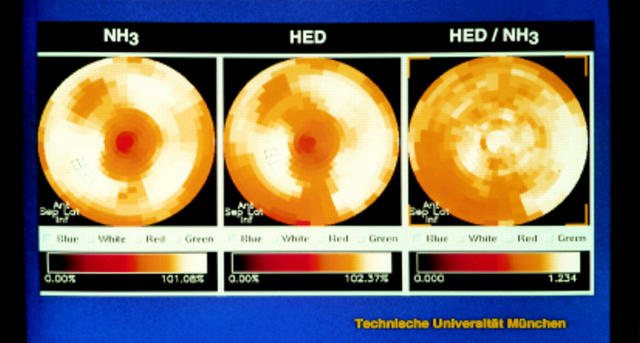
Figure 4 .
PET study of a 50 year old patient with moderate heart failure and a left ventricular ejection fraction of 30%. Blood flow images showed a normal perfusion pattern, but HED retention index was reduced by 20% compared to normal values. The regional tracer distribution appeared normal on visual inspection with only a small region of abnormal tracer retention in the distal inferior left ventricle.

