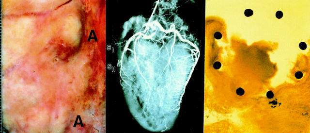
Figure 1 .
(Left) Macroscopic section of the anterior interventricular sulcus. There are two subepicardial, engorged, elastic abscesses (A) of up to 1.7 cm in diameter. (Middle) Postmortem coronary angiogram showing cessation of the contrast medium above the proximal LAD stent. The right coronary artery shows diffuse arteriosclerotic changes and restenoses of the dilated vascular section to the crux cordis. (Right) Thin section of the distal LAD stent showing eight filaments with complete, homogeneous expansion of the stent. In the outer circumferential regions of the stent filaments only remnants of the arterial wall can be seen. The major part of the vessel is destroyed by inflammation.

