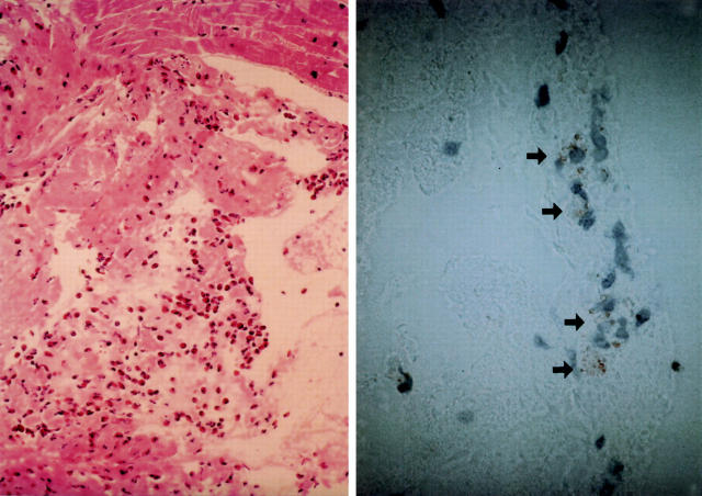
Figure 1 .
Endomyocardial biopsy (left) from the right ventricle showing slight necrosis and degeneration of myocytes, interstitial fibrosis, and infiltration of lymphocytes admixed with marked eosinophils (haematoxylin and eosin stain, original magnification × 40). Immunostaining with EG2 (arrow) (right), a monoclonal antibody directed against the secreted form of ECP, showed a great number of activated eosinophils (original magnification × 100).

