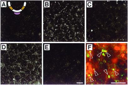
Figure 1.
Internalization of ShhN by neural plate cells in intermediate explants. The intermediate region is ochre and the notochord is mauve in the schematic cross section of the developing neural tube in A. Optical sections from the centers of explants are shown. (A) Explant incubated without ShhN and stained with polyclonal anti-Shh. (B) Explant incubated in 1 nM ShhN for 14 h and stained with polyclonal anti-Shh. (C) Explant incubated in 1 nM ShhN for 14 h and stained with anti-Shh mAb 5E1. (D) Explant incubated for 14 h in 1 nM ShhN preabsorbed with a mAb 1D4B and stained with polyclonal anti-Shh. (E) Explant incubated for 14 h in 1 nM ShhN preabsorbed with anti-Shh mAb 5E1 and stained with polyclonal anti-Shh. (F) Explant incubated for 12 h with 1 nM ShhN plus Texas Red-conjugated dextran (red) and stained with polyclonal anti-Shh (green). Open arrowheads highlight Shh+/dextran+ vesicles. The nature of the larger dextran+ structures is unclear. Both scale bars = 10 μm; the bar in E applies to A–D as well.

