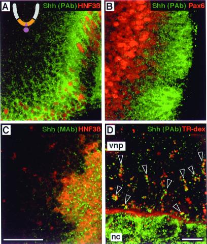
Figure 2.
Ventral neural plate cells internalize endogenous Shh. The ventral region is ochre, and the notochord and floor plate are mauve in the schematic cross section of the developing neural tube in A. (A–C) Optical sagittal sections with the floor plate to the right/lower right. (A) Explant double-labeled with polyclonal anti-Shh (green) and anti-HNF3β (red). The nuclei of HNF3β+ floor plate cells (red) are evident along the rostral-caudal axis. (B) Explant double-labeled with polyclonal anti-Shh (green) and anti-Pax6 (red). Nuclei of cells expressing low levels of Pax6 are immediately adjacent to Pax6-negative floor plate cells, and are surrounded by punctate Shh+ structures. (C) Explant double-labeled with anti-Shh mAb 5E1 (green) and anti-HNF3β (red). 5E1 detects punctate structures among HNF3β+ floor plate nuclei, presumably in secretory vesicles, with scant labeling of HNF3β-negative cells outside the floor plate. (D) An explant of ventral neural plate (vnp) with the notochord (nc) attached, from a stage before Shh expression in the floor plate, incubated with 0.5 mM leupeptin and Texas Red-conjugated dextran (red) for 6 h, then stained with polyclonal anti-Shh (green). Dextran is seen within the extracellular space between the notochord and neural plate. Open arrowheads indicate clusters of Shh+/dextran+ endocytic vesicles. Note that Shh+ structures in the notochord do not label with dextran, consistent with a biosynthetic character. Scale bars: 25 μm (A–C) and 10 μm (D).

