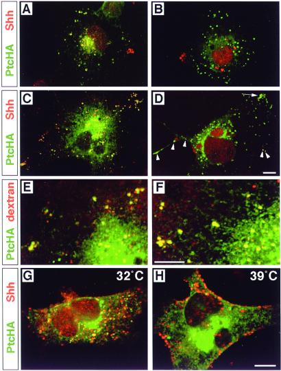
Figure 3.
Requirement of 5E1-sensitive ligand binding and dynamin activity for Ptc-mediated internalization of ShhN and ligand-independent localization of Ptc in endocytic vesicles. PtcHA immunofluorescence is shown in green and Shh immunofluorescence in red; yellow/orange indicates colocalization. (A) A single PtcHA-expressing COS cell is shown, incubated for 2 h in 1 nM ShhN pretreated with 50 μg/ml mAb 1D4B. (B) A PtcHA-expressing COS cell incubated for 2 h with 1 nM ShhN pretreated with 50 μg/ml (≈275 nM) anti-Shh mAb 5E1. No ShhN is internalized. Some red autofluorescent granules are near the nucleus, as well as debris at the left margin of the cell. (C) A single PtcHA-expressing COS cell is shown, incubated in 1 nM ShhN for two hrs, followed by detection with polyclonal anti-Shh. (D) A PtcHA-expressing cell incubated in 1 nM ShhN for two hrs, followed by detection with 5E1. Internalized ShhN is detected strongly as yellow signal in only a few isolated vesicles (arrowheads), and weakly in several within a cluster (arrow). Large red autofluorescent granules are near the nucleus. (E) A PtcHA-expressing COS cell incubated for 2 h in fluorescent dextran (red). PtcHA+/dextran+ vesicles (yellow) are evident in filopodia as well as the perinuclear region. (F) A PtcHA-expressing cell incubated in both 1 nM ShhN and fluorescent dextran for 2 h. PtcHA+/dextran+ vesicles (yellow) are evident in a pattern similar to that in E. (G) A HeLa cell overexpressing dynamints incubated in 1 nM ShhN for 2 h at 32°C. Note that PtcHA+ vesicles with internalized ShhN appear throughout the cytoplasm. (H) A PtcHA+ HeLa cell overexpressing dynamints incubated in 1 nM ShhN for 2 h starting 15 min after a shift to 39°C. Shh immunofluorescence appears predominantly in large collections at the cell surface. Scale bars = 10 μm (bar in D applies to A–C; bar in F applies to E; and bar in H applies to G).

