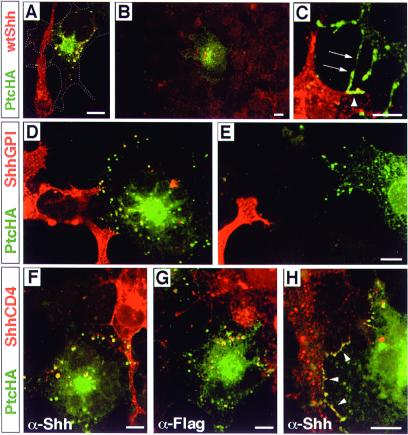
Figure 4.
Internalization of membrane-bound forms of Shh. Throughout, the Shh form is visualized in red and PtcHA in green; colocalization is seen as yellow. (A) A PtcHA-expressing cell, whose borders are indicated by the dashed line, is shown contacting a wtShh-expressing cell. PtcHA immunofluorescence is not observed where the filopodium of the wtShh-expressing cell crosses the PtcHA-expressing cell, yet the cell contains multiple PtcHA+/Shh+ vesicles. (B) A PtcHA-expressing cell not in contact with wtShh-expressing cells shows Shh+/PtcHA+ vesicles. The PtcHA+ cell is surrounded by untransfected cells in a near-confluent field; the nearest wtShh-expressing cell is out of the field of view. Nonspecific background stippling and autofluorescence is evident in untransfected cells. (C) PtcHA immunoreactivity colocalizing with wtShh at points of contact between cells at 4°C. Processes of a PtcHA-expressing cell (arrows) are shown contacting a filopodium (arrowheads) of a wtShh-expressing cell. Thirty-three cell pairs were analyzed, which included 10 pairs that were not in direct contact; all showed evidence of Shh internalization. (D) A PtcHA-expressing cell is shown in contact with filopodia of two ShhGPI-expressing cells (lower left). Shh+/PtcHA+ vesicles are yellow. (E) Shh immunoreactivity is not observed within a PtcHA+ cell that is in close proximity, but not in contact with a filopodium of a ShhGPI-expressing cell. For ShhGPI, 24 cell pairs in contact were analyzed and all showed evidence of ShhGPI internalization. (F) A PtcHA-expressing cell is shown in contact with a ShhCD4-expressing cell (upper right). Shh+/PtcHA+ vesicles are yellow. (G) Two PtcHA-expressing cells are shown in contact with a cell expressing ShhCD4F (upper right), which contains a cytoplasmic Flag epitope. Flag+/PtcHA+ vesicles are yellow. (H) Colocalization of PtcHA and ShhCD4 at points of contact (arrowheads) between cells was observed frequently in cells incubated at 37°C. For ShhCD4, a total of 32 cell pairs in contact were analyzed, and all showed evidence of ShhCD4 internalization. In control experiments, untransfected KNRK cells showed no evidence of Shh internalization when cocultured with COS cells expressing any of the Shh forms. Scale bars = 10 μm (the bar in E applies to D as well).

