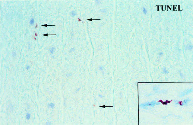
Figure 3 .
High power magnification from a TUNEL stained section of a segment of a human aorta with CMD at some distance from the dissection plane. There are several small TUNEL positive particles (arrows) probably corresponding to the apoptotic bodies shown in fig 6C. Inset: double labeling showing medial VSMCs with a positive TUNEL stain in the nucleus (brown reaction product) and cytoplasmic staining for α-actin (blue reaction product, APAAP method). One TUNEL positive nucleus shows the typical fragmentation which is a morphological feature of apoptosis.

