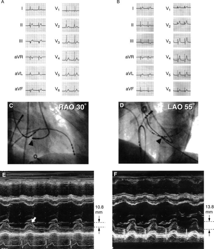
Figure 2 .

A patient with left posterior septal accessory pathway. (A) and (B), ECG recording before and after ablation. On the basis of the Oklahoma ECG algorithm, all three cardiologists assessed the location to be left posterior septum; on the basis of the St George's algorithm, the location was left posterior. (C) Right anterior view and (D) left anterior view: catheter position (indicated by arrowheads) for radiofrequency ablation. The catheter was positioned at the left posterior of the septum. (E) and (F): M mode echocardiography tracings before and after the ablation. Notch movement of the left ventricular posterior wall systolic motion can been seen in (E) (arrow) but the notch is not seen in (F). Reduced left ventricular posterior wall systolic wall motion (10.8 mm) can be seen before ablation. After ablation, the systolic motion was normalised (13.8 mm).
