Figure 1.
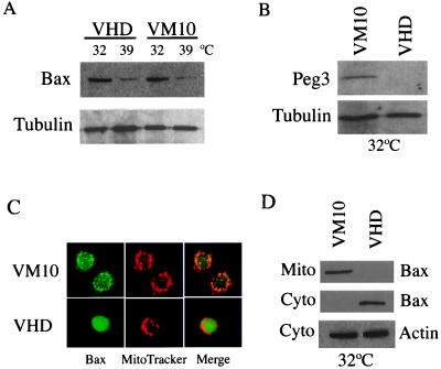
The VHD and VM10 cells showed similar Bax protein levels but distinct subcellular localization. (A) Expression of Bax protein in VHD and VM10 cells. Whole cell extracts were prepared from VHD and VM10 cells at 32°C (24 h after temperature shift) and 39°C, respectively. The Bax protein was detected by Western Blotting using a monoclonal antibody against Bax. The same blot was stripped and probed with an anti-α-tubulin antibody for loading control. Quantitation was done on a phosphorimager (Molecular Dynamics). (B) Induction of Peg3/Pw1 protein in VM10 cells at 32°C. Protein extracts isolated from VHD and VM10 cells at 32°C were blotted with an anti-Peg3/Pw1 antibody. Peg3/Pw1 is only expressed in VM10 cells at 32°C. (C) Immunostaining of the Bax protein in VHD and VM10 cells at 32°C. The mitochondria were stained with MitoTracker Red CMX Ros (red), and the Bax protein was stained with the monoclonal antibody and visualized with an FITC-conjugated secondary antibody (green). A representation of the staining pattern is shown. (D) Subcellular fractionation of Bax protein in VHD and VM10 cells. Cytosolic and mitochondria extracts were prepared from VHD and VM10 cells at 32°C. The protein concentration was measured using a protein assay kit (Bio-Rad). An equal amount of protein was loaded onto the gel. The Bax protein was detected by Western blots, and cytosolic β-actin was used as a control.

