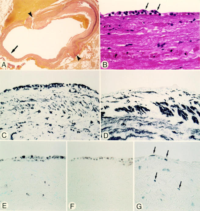
Figure 1 .

Micrographs of an anastomotic site, taken two days after grafting (patient 1). Panels A-G are serial sections. (A) Elastic tissue stain. The site of anastomosis, indicated by arrowheads, with the right coronary artery. The luminal surface of the vein graft shows a cellular response. Details of the cellular response, indicated by the arrow, are shown in panels B-G. (B) Haematoxylin and eosin stain. At the luminal surface endothelial cells have been denuded. The earliest cellular response of the grafts is demonstrated by an accumulation of polymorphonuclear leucocytes and mononuclear round cells, amid a fibrin-platelet thrombus, partially covered by spindle shaped cells (arrows). (C) Vimentin stain. Spindle shaped cells and round cells at the response site are positive. (D) HHF-35. The spindle shaped cells and round cells are negative. (E) HAM-56. Some round cells stain with this macrophage marker. None of the spindle shaped cells at the luminal site stain positive. (F) UCHL-1. Some small round cells stain with this T lymphocyte marker. (G) PCNA. Positive cells are seen at the site of cellular response and in the adjacent pre-existent media. Original magnification: (A) × 18; (B) × 580; (C-G) × 360.
