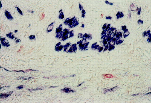
Figure 2 .
Double immunostaining with HHF-35 (blue) and PCNA (red) of an anastomotic site, two days after grafting (patient 1). The media contains a few PCNA positive cells (red), which lack staining for actin and most likely represent dedifferentiated SMCs. Most SMCs are actin positive (blue). Original magnification × 720.

