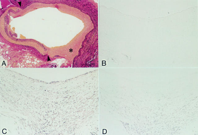
Figure 4 .
Micrographs of an anastomotic site, 33 days after grafting (patient 6). Panels A-D represent serial sections. (A) Elastic tissue stain. The anastomotic site (arrowheads) to the obtuse marginal branch shows distinct neointimal proliferation of the vein graft (asterisk). (B) HAM-56. No positivity for macrophages in the neointima. (C) HHF-35. Spindle shaped cells in the neointima stain positive. (D) CGA-7. Spindle shaped cells within the deeper layers of the neointima stain positive, but those closer to the luminal site are negative suggesting that these cells have not yet fully differentiated. The anti-vWf antibody was negative at the luminal side (not shown). Original magnification: (A) × 36; (B-D) × 145.

