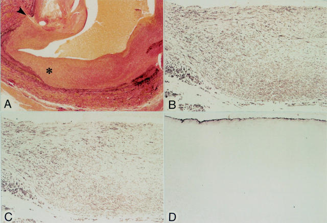
Figure 5 .
Micrographs of an anastomotic site, nine months after grafting (patient 8). Panels A-D represent serial sections. (A) Elastic tissue stain. Note distinct neointimal tissue at the anastomotic site (arrowhead). The neointima at the site of the asterisk is shown at higher magnification in panels B-D. (B) HHF-35. Spindle shaped cells in the neointima are positive. (C) CGA-7. Most spindle shaped cells are positive suggesting that most cells have the phenotype of fully differentiated SMCs (compare to panel B). (D) Anti-vWf. Regenerated endothelial cells line the luminal surface. Original magnification: (A) × 30; (B-D) × 90.

