Figure 6.
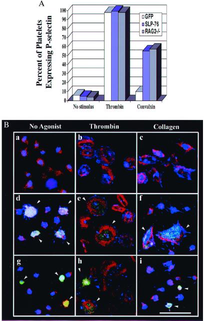
In vivo reconstitution of SLP-76 restores platelet function in SLP-76−/− mice. (A) Platelets were isolated from the nonmanipulated RAG2−/− mice or retrovirally reconstituted mice and left either untreated or stimulated with thrombin or convulxin for 10 min and then analyzed by flow cytometry for P-selectin expression. For the bone marrow reconstituted mice, gates were set to analyze only GFP-positive (transduced) cells. Similar results were seen in three independent experiments. (B) Platelets from nonmanipulated RAG2−/− mice or mice reconstituted with manipulated bone marrow were incubated for 45 min on fibrinogen-coated coverslips in the absence (a, d, and g) or presence of 0.5 unit/ml thrombin (b, e, and h), or 30 μg/ml collagen (c, f, and i). Cells were then fixed, permeabilized, and stained for phosphotyrosine (blue) and F-actin (red). Each field with platelets from manipulated mice contains a mixture of GFP-positive (transduced) and GFP-negative (nontransduced) platelets. Arrows indicate GFP-positive cells. Platelets from nonmanipulated RAG2−/− mice are shown in a-c, and d-f show platelets from a RAG2−/− mouse reconstituted with SLP-76−/− bone marrow expressing SLP-76 and GFP. (g–i) Platelets from a RAG2−/− mouse reconstituted with SLP-76−/− bone marrow expressing GFP. All panels are the same magnification. (Bar = 10 μm.)

