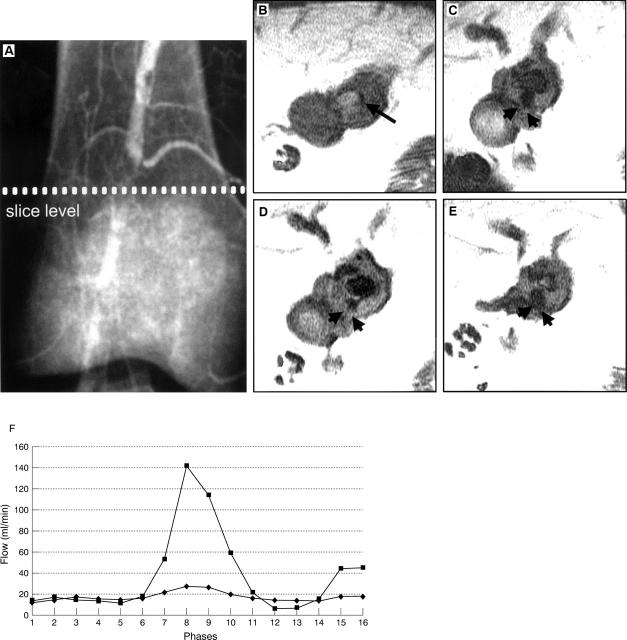
Figure 2 .
(A) Conventional angiogram of the right popliteal artery in patient 1 with a 90% stenosis, who subsequently underwent angioplasty. High resolution magnetic resonance images at the level of the stenosis show cross sections of the artery before angioplasty (B), at one week (C), at one month (D), and at three months (E). The vessel lumen appears black on all images and is shown as a small slit preangioplasty (long arrow). Angioplasty produces extensive fissuring which heals with restenosis but fissuring remains (arrowheads C-E). (F) Flow (ml/min) during 16 phases of the cardiac cycle cranial to the stenosis before angioplasty (♦) and at one week after angioplasty (▪).

