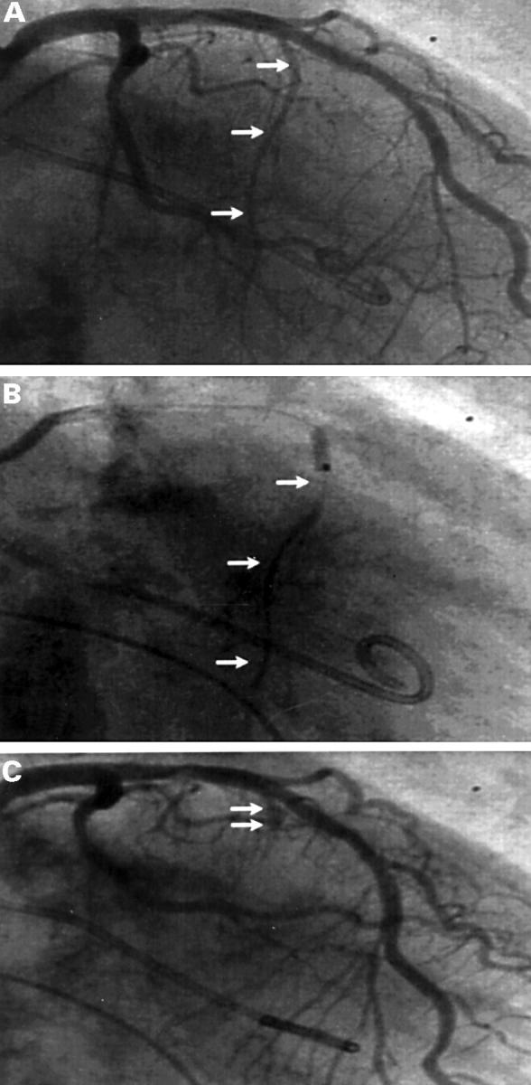
Figure 1 .

Coronary angiograms. (A) Identification of the target vessel in right anterior oblique position (arrows). (B) Injection of contrast dye to define the perfusion area and to exclude reflux into the left anterior descending coronary artery. (C) Final visualisation of the vessel stump after completed PTSMA.
