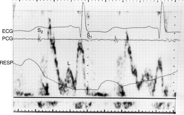
Figure 3 .
Pulsed wave Doppler velocity tracing with simultaneous ECG, phonocardiogram (PCG), and respirometer (RESP) in a patient assessed late after the Fontan operation. Mid diastolic antegrade flow (L) is shown, which occurs between the early diastolic and atrial systolic flow. Isovolumic relaxation flow, present also in this patient, is faintly shown. S1, first heart sound; S2, second heart sound.

