
Figure 1 .
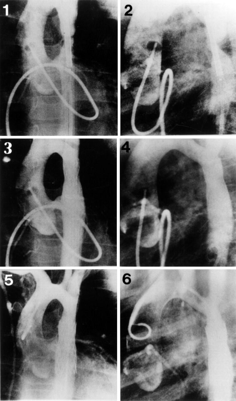
Antegrade ascending aortograms in the anteroposterior (1) and lateral (2) projections, showing the position of a stent across the coarctation before expansion. Repeat anteroposterior (3) and lateral (4) aortogram projections showed an aortic diameter of 13 mm at the stented segment and good stent position. Retrograde anteroposterior (5) and lateral (6) ascending aortogram projections 36 months later showed no evidence of restenosis and good stent position without fracture or displacement (patient 2, tables 1 and 2).