
Figure 3 .
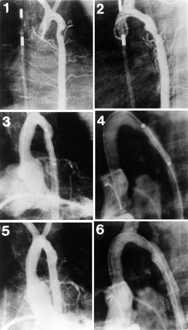
Ascending aortograms in the anteroposterior (1) and lateral (2) projections, revealing a discrete recoarctation in a 4 month old infant after a left subclavian flap operation. Left ventriculography in the anteroposterior (3) and lateral (4) projections show the position of a stent across the coarctation before expansion. Left ventriculography in the anteroposterior (5) and lateral (6) projections show a reasonable increase in coarctation diameter following stent placement (patient 1, tables 1 and 2).