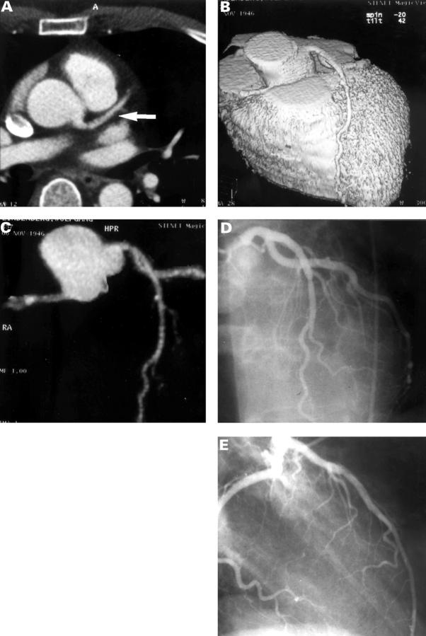
Figure 1 .
Contrast enhanced EBCT study in a 36 year old male patient after anterior myocardial infarction, without significant stenoses on invasive coronary angiography. (A) Contrast enhanced tomogram at the level of the aortic root shows the left main and left anterior descending coronary artery (arrow). (B) Three dimensional reconstruction of the heart showing left anterior descending coronary artery without significant stenosis. The surface reconstruction is thresholded to show only the contrast enhanced lumen of the coronary arteries. (C) Maximum intensity projection of the coronary arteries showing left anterior descending coronary artery without stenosis. (D, E) Invasive coronary angiography: absence of significant stenoses in the left anterior descending coronary artery after anterior myocardial infarction.

