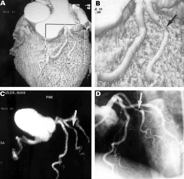
Figure 2 .
Thirty nine year old male patient after anterolateral myocardial infarction. (A, B) Surface reconstruction shows a high grade stenosis in a large diagonal branch (arrow). (C) Maximum intensity projection of the coronary arteries (arrow: stenosis of diagonal branch). (D) Invasive coronary angiography confirms high grade stenosis of the diagonal branch (arrow).

