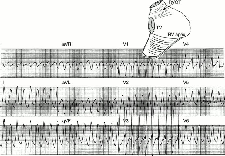
Figure 1: .

Idiopathic right ventricular outflow tract tachycardia. The 12 lead ECG shows tachycardia with a left bundle branch block, configuration and frontal plane axis directed inferiorly. The schematic at the upper right shows the right ventricle viewed from the right anterior oblique position with the free wall of the ventricle folded down. The location of the tachycardia in the right ventricular outflow tract (RVOT) is indicated with an arrow. TV, tricuspid valve; RV, right ventricle.
