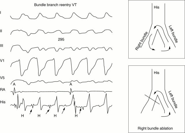
Figure 3: .
Bundle branch re-entry tachycardia. The left hand panel shows bundle branch re-entry tachycardia initiated in the electrophysiology laboratory. From the top are surface ECG leads and intracardiac recordings from the right atrium (RA) and His bundle position (His). VT has a left bundle branch block configuration and cycle length of 295 ms. Atrioventricular dissociation is evident in the right atrial recording (RA). A His bundle deflection (arrows) precedes each QRS indicating that the His-Purkinje system is closely linked to the tachycardia. The schematic in the right hand panels illustrates the mechanism. The wavefront circulates down the right bundle, through the interventricular septum, and up the left bundle (top panel). Ablation of the right bundle branch interrupts the circuit (bottom panel).

